.png)
How Pigmentation Forms Under Skin: A Clear Guide
- chevonne stewart
- Jun 14
- 8 min read


Skin pigmentation forms through a biological process called melanogenesis, where specialized cells called melanocytes produce melanin pigment and transfer it to surrounding skin cells. This process is your skin’s primary defense against ultraviolet damage. Melanocytes sit in the deepest layer of the epidermis and work alongside keratinocytes in what scientists call the epidermal melanin unit. Two key enzymes drive the process: tyrosinase initiates the conversion of L-tyrosine into melanin, while related enzymes determine whether your skin produces eumelanin (brown-black) or pheomelanin (red-yellow). Understanding this biology is the first step toward understanding why pigmentation changes and how to address it.
How pigmentation forms under skin at the cellular level
Melanogenesis is a multi-step enzymatic process that begins with a single amino acid. Tyrosinase converts L-tyrosine into DOPA, then into dopaquinone, which branches into either eumelanin or pheomelanin depending on the presence of cysteine. This enzymatic cascade is the core of melanin synthesis and determines both the color and intensity of your skin tone.
Melanin does not float freely inside cells. It is packaged inside organelles called melanosomes, which mature through four distinct stages before they are ready for transfer. As melanosomes mature, they move from the center of the melanocyte outward along the cell’s extensions, called dendrites, toward neighboring keratinocytes. This transport relies on a precise cytoskeletal mechanism involving both microtubules and actin filaments. When those structures are disrupted, melanin distribution fails and UV protection is compromised.

Once keratinocytes absorb the melanosomes, something remarkable happens. The melanosomes cluster above the keratinocyte nucleus, forming what researchers call a supranuclear melanin cap. This cap acts like a physical shield, absorbing UV radiation before it can damage DNA. Melanosomes localize predominantly over nuclei in basal keratinocytes, providing direct DNA protection. The efficiency of this cap determines how well your skin handles sun exposure.
The steps in this process, in order, are:
L-tyrosine is converted to DOPA by tyrosinase inside the melanosome
DOPA oxidizes into dopaquinone, the branching point for melanin type
Melanosomes mature through four stages and load with melanin
Mature melanosomes travel along microtubules and actin filaments to dendrite tips
Melanosomes transfer into keratinocytes via direct injection or vesicle release
Keratinocytes position melanosomes above their nuclei as protective caps
Pro Tip: The number of melanosomes transferred, not just the number of melanocytes, determines how dark your skin appears. This is why two people with similar melanocyte counts can have very different skin tones.
What triggers pigmentation to develop in your skin?
UV radiation is the most studied trigger for increased melanin production. When UV hits the skin, keratinocytes release alpha-melanocyte-stimulating hormone (α-MSH), which binds to the MC1R receptor on melanocytes. This activates the cAMP/PKA signaling pathway, which in turn activates MITF, the master transcription factor for melanogenesis. UV exposure triggers this entire cascade, increasing melanin synthesis as a photoprotective response. The result is the tan you see days after sun exposure.
Visible light and near-infrared radiation also contribute to pigmentation, and this is where many people are surprised. Blue light activates the OPN3 receptor in melanocytes, triggering calcium and MAPK signaling that increases both MITF activity and tyrosinase production. Blue light activates OPN3, producing pigmentation that often persists longer than UV-induced changes. Screen exposure and indoor lighting are real, if modest, contributors to pigmentation in susceptible individuals.
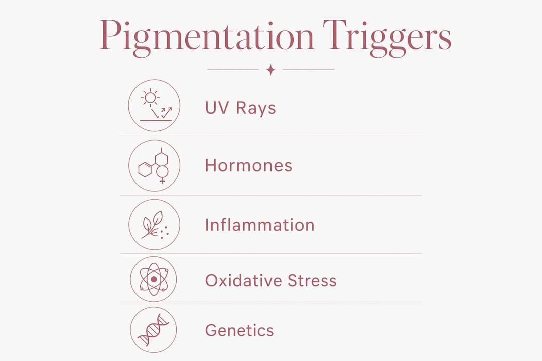
Several other triggers also influence how skin pigmentation develops:
Inflammation: Acne, eczema, and skin injuries trigger the release of prostaglandins and cytokines that upregulate melanogenesis through the MITF pathway
Hormones: Estrogen and progesterone increase melanocyte sensitivity, which is why pregnancy and contraceptive use are strongly linked to melasma
Oxidative stress: Free radicals generated by pollution or UV exposure activate NF-κB signaling, further amplifying melanin output
Genetics: Variants in genes controlling tyrosinase activity and MC1R receptor sensitivity determine how strongly your melanocytes respond to any given trigger
The MITF transcription factor sits at the center of all these pathways. Signals from UV, hormones, inflammation, and oxidative stress all converge on MITF. This convergence explains why pigmentation disorders are so difficult to treat with a single approach and why they tend to relapse when triggers are not fully controlled.
How do different pigmentation types form and persist?
Not all pigmentation forms the same way. The mechanism behind each type determines how long it lasts and how deep it sits in the skin.
Pigmentation Type | Primary Trigger | Skin Layer Affected | Persistence |
Sun-induced hyperpigmentation | UV radiation via α-MSH/MC1R | Epidermal | Weeks to months |
Melasma | Hormones, UV, genetics, blue light | Epidermal and dermal | Chronic, relapsing |
Post-inflammatory hyperpigmentation | Skin injury or inflammation | Epidermal (sometimes dermal) | Weeks to months after healing |
Dermal pigmentation | Melanin deposited in dermis | Dermal | Long-lasting, treatment-resistant |
Melasma is the most complex type. It is associated with female sex, pregnancy, and hormonal contraceptive use, and it appears on sun-exposed facial areas. What makes melasma particularly stubborn is that multiple triggers operate simultaneously. Hormones prime the melanocytes, UV and blue light activate them, and genetic predisposition amplifies the response. Even after one trigger is removed, the others sustain the pigmentation. This is why melasma has a chronic, relapsing character that frustrates both patients and clinicians.
Post-inflammatory hyperpigmentation (PIH) forms after skin injury or inflammation, including acne, eczema, or cosmetic procedures. The inflammatory process stimulates melanocytes to overproduce melanin, which then loads into keratinocytes. Here is the detail most people miss: PIH pigmentation becomes visible only as melanin-laden keratinocytes migrate upward over 28–40 days. The dark spot you see weeks after a pimple heals is not new pigment. It is old pigment arriving at the skin surface on schedule.
Dermal pigmentation is the most treatment-resistant form. When melanin deposits in the dermis rather than the epidermis, it sits below the reach of most topical treatments. This happens when the epidermal barrier is severely disrupted, allowing melanin to fall into deeper tissue. Clinically, hyperpigmentation can involve epidermal or dermal compartments, and the depth of pigmentation directly affects treatment response and prognosis. A Wood lamp examination helps clinicians identify which layer is involved before choosing a treatment path.
Why does pigmentation vary so much between individuals?
Skin color differences arise not from the number of melanocytes, which is roughly equal across all skin tones, but from how those melanocytes behave. The key variables are melanosome size, packaging, and the ratio of eumelanin to pheomelanin.
In darker skin tones, melanosomes are larger, more numerous, and distributed individually throughout the keratinocyte. In lighter skin tones, melanosomes are smaller and grouped in clusters. This difference in melanosome size and distribution explains why darker skin provides stronger natural UV protection and why it is also more prone to visible post-inflammatory pigmentation after injury.
The eumelanin-to-pheomelanin ratio also matters significantly. Eumelanin is photoprotective and produces brown to black tones. Pheomelanin is less stable, produces red to yellow tones, and generates free radicals when exposed to UV. People with higher pheomelanin ratios, typically those with fair or red-toned skin, experience more UV-induced oxidative stress per unit of sun exposure. Tyrosinase activity and genetic regulation, including MC1R variants, control which type of melanin dominates.
Pro Tip: If you notice that your skin darkens significantly after even mild sun exposure, your melanocytes likely have high tyrosinase activity or a sensitive MC1R receptor. This makes consistent daily sun protection more important for you than for someone whose skin responds less readily. Explore pigmentation best practices for guidance tailored to your skin type.
Small differences in melanosome maturation and trafficking create variation in pigmentation even when melanocyte activation is similar. This is why two people with the same skin tone can respond very differently to the same UV exposure or inflammatory event. The diversity in pigmentation patterns among individuals comes down to organelle-level differences, not just surface-level skin type.
Key takeaways
Skin pigmentation forms through melanogenesis, a multi-step process controlled by melanocytes, tyrosinase, MITF signaling, and melanosome transfer to keratinocytes, with UV, hormones, and inflammation as the primary triggers.
Point | Details |
Melanogenesis is the core process | Melanocytes convert L-tyrosine into melanin via tyrosinase and package it into melanosomes for transfer. |
MITF controls all major triggers | UV, hormones, inflammation, and blue light all converge on MITF, making pigmentation multifactorial by nature. |
Pigmentation visibility is delayed | Melanin-laden keratinocytes take 28–40 days to migrate to the surface, so spots appear weeks after the trigger. |
Melanosome behavior drives skin tone | Melanosome size, distribution, and eumelanin-to-pheomelanin ratio determine individual skin color variation. |
Dermal pigmentation resists treatment | Melanin deposited in the dermis sits below topical reach and requires professional clinical assessment. |
What 15 years of treating pigmentation has taught me
The most common misconception I see in clinic is that pigmentation is simply “too much melanin.” That framing leads people to reach for brightening serums and expect fast results. The biology tells a different story.
Pigmentation is a system response, not a single event. When I assess a client with melasma, I am looking at the interaction of hormonal history, sun exposure habits, skin barrier integrity, and genetic predisposition all at once. Treating only one factor while the others remain active is why so many people cycle through treatments without lasting improvement. The multifactorial nature of melasma is not a clinical footnote. It is the central clinical challenge.
The 28–40 day keratinocyte migration timeline is also something I wish more clients understood before they start treatment. When you begin addressing pigmentation, you will not see results in a week. The melanin already loaded into keratinocytes has to travel to the surface and shed before clearer skin is visible. Clients who understand this timeline stay the course. Those who do not often abandon effective treatments too early.
What I find most clinically interesting is how blue light and near-infrared radiation contribute to pigmentation persistence, especially in clients who work indoors under artificial lighting or spend significant time on screens. This is a relatively recent area of research, and it changes how I counsel clients on daily protection. Sunscreen alone is not always enough. Antioxidant support and barrier care matter too.
Pigmentation is not a flaw to be erased. It is a biological response that, once understood, becomes far more manageable. Understanding why your skin responds the way it does is not just reassuring. It is the foundation of any treatment plan that actually works.
— chevonne
Ready to address your pigmentation with professional support?
If this breakdown of pigmentation biology resonates with what you are experiencing in your own skin, you are not alone. At Fundamentalskin, Chevonne works with women dealing with exactly these concerns, combining clinical expertise with targeted treatments designed for real, lasting results.

Fundamentalskin offers three professional peel options to support pigmentation management. The Larimedical Peel targets melanin overproduction at the cellular level. The Biomimetic Peel with LED Therapy combines exfoliation with light-based support for more complex pigmentation. The Synergie Peel offers a gentler entry point for sensitive or reactive skin. All treatments are non-invasive, backed by before-and-after results, and tailored to your unique skin profile. BOOK NOW to start your consultation with Chevonne.
FAQ
What is melanogenesis and why does it matter?
Melanogenesis is the biological process by which melanocytes produce melanin pigment inside organelles called melanosomes and transfer them to keratinocytes. It is the foundation of all skin pigmentation, both natural skin tone and pigmentation disorders.
Why does a dark spot appear weeks after a pimple heals?
Post-inflammatory hyperpigmentation becomes visible as melanin-laden keratinocytes migrate to the skin surface over 28–40 days after the original inflammation. The delay is a normal part of the skin cell cycle, not a sign that pigmentation is worsening.
Does blue light from screens cause skin pigmentation?
Blue light activates the OPN3 receptor in melanocytes, triggering signaling pathways that increase tyrosinase activity and melanin production. The resulting pigmentation can persist longer than UV-induced changes, making daily antioxidant protection relevant even indoors.
Why does melasma keep coming back after treatment?
Melasma is driven by multiple simultaneous triggers including hormones, UV, blue light, and genetic predisposition, all converging on the MITF transcription factor. Removing one trigger while others remain active allows melanocyte stimulation to continue, causing relapse.
Is skin tone determined by the number of melanocytes?
Skin tone is determined not by melanocyte count, which is roughly equal across all skin tones, but by melanosome size, distribution within keratinocytes, and the ratio of eumelanin to pheomelanin. You can explore treatment options compared to understand how these differences affect clinical care.
Recommended
Comments