.png)
Freckles vs. Sunspots: How to Tell Them Apart Safely
- chevonne stewart
- Aug 17
- 11 min read

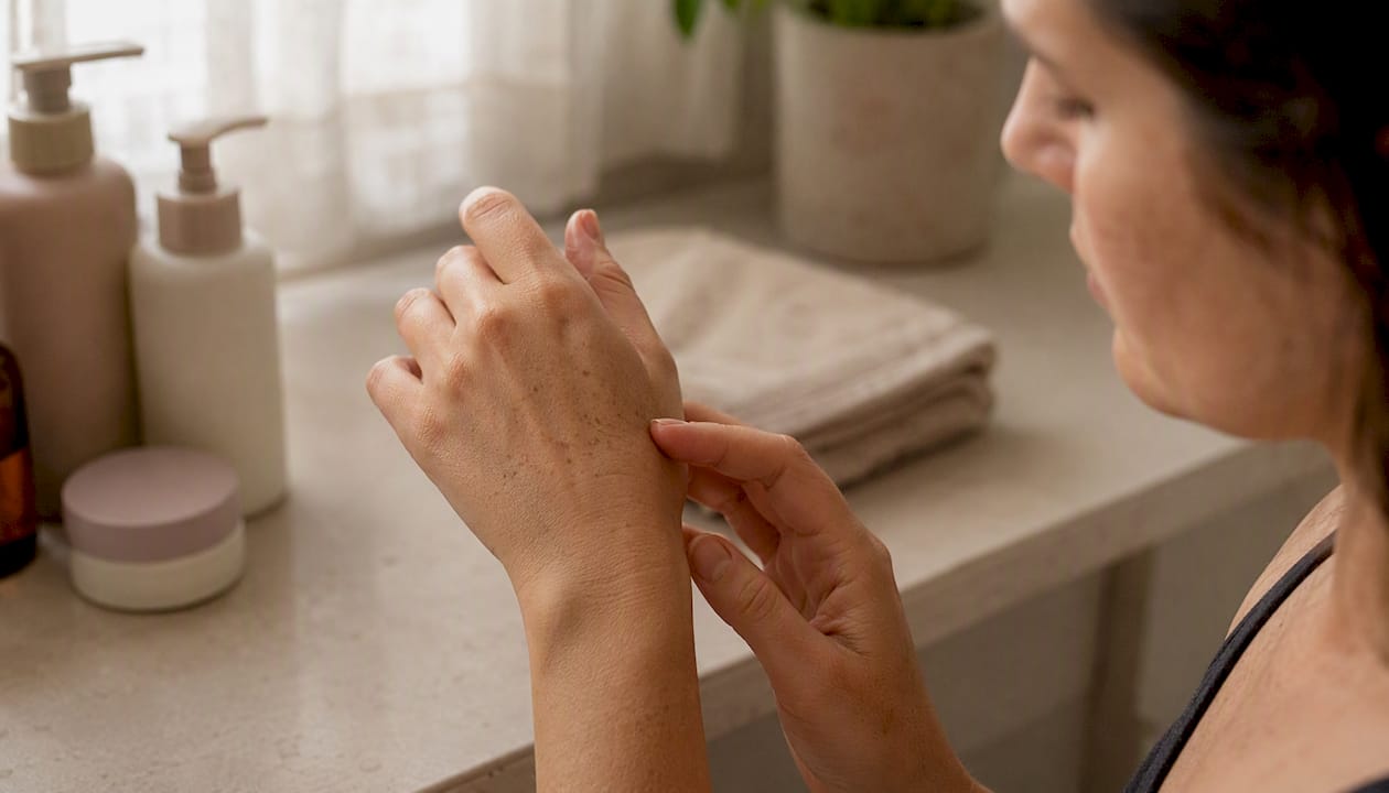
A freckle is a genetic, sun-reactive mark that shows up young, darkens in summer, and fades in winter. A sunspot, medically called a solar lentigo, is a flat patch of cumulative UV damage that shows up later in life and never really goes away. That single distinction, genetics-plus-seasonality versus decades of sun exposure, is the fastest way to tell them apart before you even look at size or color.
Here’s the quick visual rundown you can use in the mirror right now:
Size: Freckles usually stay under 5 mm; sunspots can grow to nearly 1.5 cm.
Age of onset: Freckles appear in early childhood; sunspots typically show up after age 40.
Seasonality: Freckles darken in summer and fade in winter; sunspots stay put year-round.
Color: Freckles range from light red to brown; sunspots tend toward a more uniform tan to dark brown.
Cancer risk: Both are benign on their own, but either can sit alongside precancerous lesions if the texture changes.
Pro Tip: If a spot suddenly feels rough, scaly, or starts to itch or bleed, don’t wait for your next scheduled skin check. Book a clinical evaluation that week, not “eventually.”
Key Takeaways
Freckles are genetic, seasonal, and appear young; sunspots are cumulative sun damage that persists year-round and shows up later in life, and texture change is the signal that always warrants a clinical check.
Point | Details |
Core visual difference | Freckles fade in winter and stay small; sunspots persist year-round and can grow larger. |
Age pattern matters | Freckles start in early childhood; sunspots typically appear after age 40. |
Texture is the red flag | Rough, scaly, or bleeding spots need prompt evaluation regardless of size or color. |
Prevention beats correction | Daily broad-spectrum SPF prevents new spots since treatment doesn’t erase existing sun damage. |
Clinic option for pigmentation | Fundamentalskin’s Larimedical Biomimetic Peel targets uneven tone from sun damage without downtime. |
Table of Contents
What Is the Difference Between Freckles and Sunspots?
The clinical difference comes down to origin. Freckles, known medically as ephelides, are a genetic trait triggered by sun exposure. Sunspots, or solar lentigines, are a direct result of accumulated ultraviolet damage over years, sometimes decades, of unprotected sun exposure. One is written into your DNA and lit up by summer. The other is a slow-building consequence of UV history that doesn’t reverse itself when the weather cools.
This is why a nine-year-old can have freckles across her nose that vanish by January, while her grandmother has a sunspot on the back of her hand that looks the same in July as it does in December. Same brown pigment, completely different biological story. Understanding that story matters, because it changes what you should expect from any treatment and how seriously you should monitor a given mark.
What a Freckle (Ephelide) Actually Is
A freckle, clinically called an ephelide, is a small, flat pigment spot that forms when genetics and UV exposure combine. People with the MC1R gene variant, commonly associated with lighter skin, red or blond hair, and light eyes, are far more likely to develop them. Cleveland Clinic describes freckles as ranging in color from light red to brown, most often appearing on sun-exposed areas like the face, shoulders, arms, and chest.
Freckles behave predictably:
They typically appear in early childhood.
They measure roughly 1 to 5 mm, smaller than most sunspots.
They darken noticeably with summer sun exposure and lighten in winter months.
They can fade with age in some people, though not universally.
They cluster in sun-exposed zones rather than appearing as a single isolated patch.
A freckle is benign by definition. But its presence signals something worth paying attention to: a higher baseline sensitivity to UV radiation. If your skin freckles easily, your lifetime skin cancer risk trends higher than someone whose skin tans evenly without spotting, which makes daily sun protection more than a cosmetic habit for you.
What a Sunspot (Solar Lentigo) Actually Is
A sunspot, or solar lentigo, is a flat, pigment-only patch that forms after years of cumulative UV exposure rather than a single bad sunburn. Mayo Clinic notes that these are typically flat, round or oval, and darker or larger than the surrounding skin, most commonly appearing on the face, hands, shoulders, and forearms, the areas that catch the most sun over a lifetime.
Sunspots have their own distinct pattern:
They usually emerge later in life, though heavy sun exposure earlier in life can push that timeline younger.
They can reach up to roughly 1.5 cm, noticeably larger than the typical freckle.
They persist year-round and don’t fade in winter the way freckles do.
Their color tends toward a more consistent tan, brown, or occasionally near-black shade.
They’re often mistaken for “age spots,” which is the same lesion under a more casual name.
Here’s where it gets more clinically important: not every rough or scaly patch that looks like a sunspot actually is one. Actinic keratoses, a separate and precancerous condition caused by the same UV damage, can resemble sunspots at a glance but feel distinctly different to the touch. Texture is the tell, and it’s the detail most people overlook when they’re just eyeballing a mirror.
Freckles vs. Sunspots: A Side-by-Side Comparison
Seeing the differences laid out together makes the pattern click faster than reading them separately.
Dimension | Freckles (Ephelides) | Sunspots (Solar Lentigines) |
Typical age of onset | Early childhood | Usually after middle age |
Primary cause | Genetics (MC1R gene) plus UV exposure | Cumulative UV damage over decades |
Size and color | About 1 to 5 mm; light red to brown | Up to roughly 1.5 cm; tan to dark brown |
Seasonal behavior | Darkens in summer, fades in winter | Persists year-round, no seasonal fading |
Texture | Flat, smooth | Flat, smooth (unless actinic keratosis is present) |
Cancer or precancer risk | Benign, but signals higher UV sensitivity | Benign, but can resemble or coexist with actinic keratosis |
Common treatments | Topical fading agents, sun protection | Chemical peels, lasers, IPL, cryotherapy |
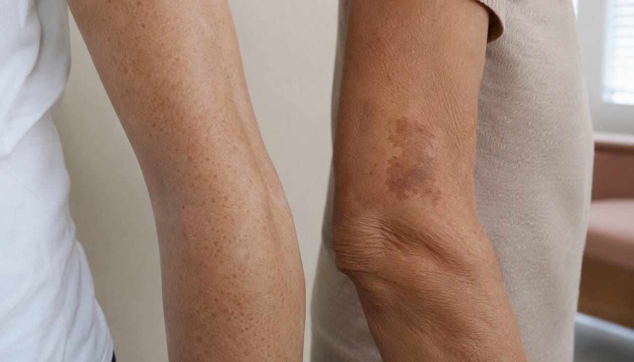
Picture two patients side by side. A seven-year-old shows up with a scatter of small freckles across her cheeks and nose bridge, brighter in July, nearly invisible by February. A 58-year-old presents with a single, persistent 1-cm brown patch on the back of his right hand that has looked identical for the past three years regardless of season. Same general appearance category, entirely different clinical read.
How to Tell Freckles and Sunspots Apart From Moles
Moles, or nevi, play by different rules than freckles and sunspots, and mixing them up is one of the most common reasons people either panic unnecessarily or ignore something that actually needs attention.
Freckles and sunspots are flat, pigment-only lesions. Moles are often raised, can have distinct borders, and grow from a cluster of pigment-producing cells rather than sitting purely on the skin’s surface. Clinicians frequently see patients confuse the two categories, which matters because moles follow a completely different monitoring protocol.
The ABCDE checklist remains the gold standard for evaluating any pigmented lesion, whether it’s a mole, freckle, or sunspot:
Asymmetry — one half doesn’t match the other.
Border — edges are irregular, notched, or blurred rather than smooth.
Color — multiple shades within one spot, or unusual colors like blue, red, or white.
Diameter — larger than about 6 mm, roughly the size of a pencil eraser.
Evolving — any change in size, shape, color, or elevation over time.
Beyond ABCDE, watch for a few extra red flags that apply specifically to sun-damage lesions:
A spot that feels scaly, rough, or crusty to the touch.
Any lesion that starts bleeding without an obvious injury.
Persistent itching in one specific spot.
Sudden or rapid growth over weeks rather than years.
Actinic keratoses are the lesion most likely to hide behind a “just a sunspot” assumption. They typically feel rough or sandpapery rather than smooth, and that texture difference is exactly why a simple touch test belongs in your monthly self-check, right alongside a visual scan and a quick photo for comparison over time.
When to See a Doctor About a Suspicious Spot
Most freckles and most sunspots never need a clinician’s attention beyond routine skin checks. But certain signs should move you from “I’ll mention it at my next appointment” to “I’m calling this week.”
Prioritize an evaluation if you notice any of the following:
A spot that has changed shape, color, or size over the past few months.
New roughness, scaliness, or crusting on a previously smooth lesion.
Bleeding, oozing, or a sore that doesn’t heal within a few weeks.
A new spot appearing after age 40 that doesn’t match your usual freckle pattern.
Persistent pain, tenderness, or itching localized to one lesion.
Any lesion meeting two or more ABCDE criteria at once.
Once you’re in front of a clinician, the diagnostic process typically follows a consistent path. It starts with a history covering your sun exposure and family history, moves into a full clinical exam, and often includes dermoscopy, a magnified, illuminated exam that lets a clinician see pigment patterns invisible to the naked eye. Photographic monitoring may follow for borderline lesions, tracking subtle changes over months. If anything still looks uncertain, a biopsy settles the question definitively.
“Suspicious or changing moles should be monitored closely or biopsied,” according to guidance from the National Cancer Institute, a standard that extends in practice to any pigmented lesion that won’t behave predictably.
The reassuring part: the overwhelming majority of biopsies on flat pigmented lesions come back as benign lentigines or ordinary freckles. But that reassurance only holds up if you actually get the changing spot checked instead of assuming it’s nothing.
Preventing New Freckles and Sunspots
Prevention beats correction every time, because no treatment fully erases the sun damage already stored in your skin. Cumulative UV exposure is what created the problem in the first place, and dermatology literature consistently links that exposure to solar lentigines and other pigmentary changes.
A handful of habits do most of the heavy lifting:
Apply a broad-spectrum SPF 20 minutes before heading outdoors, and reapply every two hours, more often if you’re swimming or sweating.
Choose a professional-grade sunscreen formulated for pigmented skin rather than a basic drugstore option, especially if you’ve already had treatment for existing spots.
Wear a wide-brim hat, UPF-rated clothing, and sunglasses when you’re outside for extended periods.
Plan outdoor activities before 10 a.m. or after 4 p.m. to avoid peak UV hours.
Skip intentional tanning, whether that’s a tanning bed or deliberate sun-worshipping, since both accelerate lentigo formation.
Take a monthly photo of any spots you’re tracking so subtle changes don’t sneak past you.
Sunscreen’s role here isn’t cosmetic polish, it’s the mechanism that actually prevents new dark spots from forming in the first place.
Pro Tip: Keep your sunscreen in the same spot as your toothbrush. Habit stacking it with something you already do twice daily is the single easiest way to make daily SPF automatic instead of optional.
How Freckles and Sunspots Can Be Lightened or Removed
Treatment options for freckles and sunspots overlap significantly, though the approach shifts depending on the lesion type and your skin tone.
Common options include:
Topical agents: Retinoids and prescription lightening creams that gradually fade pigment over weeks to months, often the safest starting point for at-home fading.
Chemical peels: Controlled exfoliation that removes pigmented surface cells, requiring a series of sessions for visible results.
Cryotherapy: Freezing the lesion with liquid nitrogen, more commonly used for isolated sunspots than widespread freckling.
Laser and IPL: Targeted light energy that breaks down pigment, generally the fastest route to visible fading for solar lentigines.
Procedural resurfacing: Deeper treatments for more stubborn or widespread pigmentation.
Actinic keratoses require an entirely different conversation. Because they carry precancer risk, they’re treated medically, cryotherapy, topical chemotherapy agents, or field therapy, not with cosmetic fading approaches meant for benign lentigines. Confusing the two categories means either over-treating a harmless freckle or under-treating a lesion that actually needs medical intervention.
Darker skin types need an extra layer of caution with any of these treatments. Aggressive peels or lasers can trigger post-inflammatory hyperpigmentation, sometimes leaving a mark darker than the one you started with.
Fading visible pigment with peels, retinoids, or lasers doesn’t erase the skin’s underlying sun-damage memory. Without consistent daily SPF afterward, new spots tend to reappear in the same UV-exposed areas within months.
That’s the piece most people skip: treatment is maintenance, not a cure. Whatever method you choose, the results only hold if daily photoprotection continues indefinitely afterward.
What to Expect From a Professional Pigmentation Consultation
A proper clinic consultation for pigmentation should never start with a treatment recommendation before it starts with a conversation. Here’s the sequence a thorough clinician follows:
Review your medical history and current medications, since some medications increase photosensitivity.
Photograph existing spots to establish a baseline for tracking change over time.
Perform dermoscopy on any lesion that looks ambiguous or has changed recently.
Discuss your goals honestly, including what’s realistic given your skin type and sun history.
Build a treatment plan around maintenance, not just a single session.
Realistic expectations matter here. Most pigmentation treatments require multiple sessions spaced weeks apart, with visible improvement building gradually rather than appearing overnight. Downtime varies by method, chemical peels typically mean a few days of visible flaking, while gentler options allow you to return to normal activity immediately.
A few practical considerations shape which approach fits:
Skin tone and sensitivity determine which treatment intensity is appropriate without risking hyperpigmentation.
The lesion’s texture, flat versus rough, determines whether cosmetic fading or medical evaluation comes first.
Your commitment to daily SPF afterward determines how long results actually last.
For clients dealing specifically with pigmentation from sun damage, the Larimedical Biomimetic Peel is one option built around this exact framework: a non-invasive approach that targets uneven tone without the downtime of more aggressive resurfacing. Pairing it with LED therapy is a common next step for clients looking to support skin recovery and even out texture alongside pigment correction, particularly useful for anyone managing both sunspots and the fine lines that tend to show up around the same age.
A Clinician’s Perspective on Freckles and Sunspots
Most of what walks through the door labeled “worrying spot” turns out to be a perfectly ordinary freckle or a benign sunspot that’s been there for years. The real work isn’t reassuring people that most pigmentation is harmless, it’s teaching them to notice the handful of signs that actually matter: a new roughness, a sudden change, a spot that itches when nothing around it does.
Prevention gets undersold constantly. People assume that once a treatment fades a spot, the problem is solved. It isn’t. The skin’s UV history doesn’t reset with a peel or a laser session. What actually protects you long-term is the boring, unglamorous habit of applying sunscreen every single day, whether or not you have a visible spot to worry about that morning.
If there’s one thing worth taking seriously from this entire conversation, it’s this: photograph anything you’re tracking, and don’t talk yourself out of an appointment because a spot “probably” hasn’t changed. Evidence-based assessment beats guesswork every time, and an individualized plan built around your actual skin, not a generic checklist, is what makes treatment last.
Book a Pigmentation Consultation That Actually Fits Your Skin
If you’ve read this far trying to figure out whether that spot on your cheek or the back of your hand deserves attention, the next step is simple: get it looked at by someone trained to tell the difference between a harmless freckle, a stubborn sunspot, and something that needs closer monitoring. Fundamentalskin’s consultations start with exactly that kind of assessment, not a generic product recommendation, before building a plan around your skin’s actual history and goals.

For clients dealing with persistent solar lentigines or uneven tone, the Biomimetic Peel + LED Therapy combines targeted pigment correction with skin recovery support, without the downtime of more aggressive resurfacing options. Book a consultation to get a personalized read on your skin before choosing a treatment path. One important note: this article, and any consultation that follows, is general guidance, not a substitute for urgent medical care. If a spot is rapidly changing, bleeding, or meets multiple ABCDE criteria, see a physician immediately rather than waiting for a cosmetic appointment.
Sources
Recommended
Comments